
Quick setup
Check the basics:
-
patient name and age
-
view (supine AP ± erect/decubitus ± erect CXR)
-
image quality/rotation/artifacts
Typical sets are supine AXR; add erect or left lateral decubitus to look for free air; many centers use an erect chest for pneumoperitoneum.1
Why are we imaging?
AXR is best for “gas, masses, bones, stones” and specific red flags such as obstruction, toxic megacolon, and perforation. CT outperforms AXR for many acute abdomens, so escalate when suspicious.2
The ABDOE system
A — Air (gas)
-
Where it should be: stomach, small bowel (a little), and colon/rectum.
-
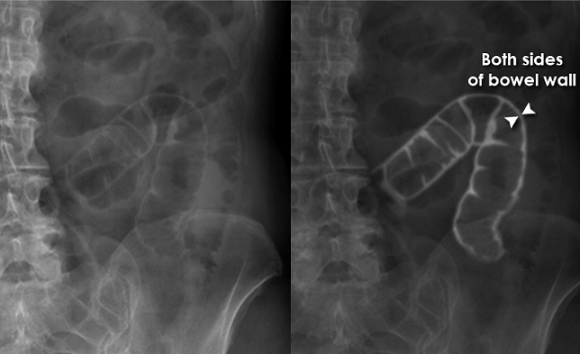
Where it shouldn’t be: free intraperitoneal air—look for the Rigler sign (double bowel wall on supine view) or subdiaphragmatic air on erect/decubitus. Treat as perforation until proven otherwise.1,3,4
B — Bowel
Location & folds:
-
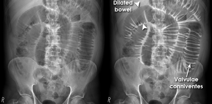
Small bowel: mostly central, with valvulae conniventes traversing the full width¹.
-
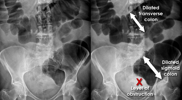
Large bowel: more peripheral, with haustra that don’t cross completely¹.
-
3–6–9 rule: small bowel <3 cm, colon <6 cm, cecum <9 cm; above this = dilated.1,3
Obstruction clues:
Small Bowel Obstruction: multiple dilated central loops, step-ladder pattern, air-fluid levels on erect, and the string-of-pearls sign (gas trapped between folds in fluid-filled loops).1,3,5
Large Bowel Obstruction: peripheral colonic dilation; check cecal size (risk of perforation when large). Sigmoid volvulus may present with a coffee-bean sign.1,3,6
Toxic megacolon: transverse colon >6 cm plus systemic toxicity.2,7,8

Hellerhoff - File:Toxisches_Megacolon_bei_Colitis_ulcerosa.jpg
C — Calcifications
-
Renal/ureteric stones: ~90% radiopaque on AXR for renal stones; many ureteric stones also visible.3,9
-
Gallstones: only ~10–15% radiopaque on AXR; often incidental.3,10
-
Vessels: curvilinear aortic wall calcification may indicate AAA—AXR is not for sizing, so use US or CT for measurement.11
-
Bones: always scan ribs, spine, pelvis, and hips for fractures or lesions.

Aslam S, Renal calculi. Case study, Radiopaedia.org
D — Devices & foreign bodies
Check for:
-
NG tubes, stents, clips, IUDs, surgical staples, jewelry, and ECG leads
-
NG tube should follow the expected path to the stomach12

E — Everything else (organs & soft tissues)
Trace liver/spleen outlines; look for organomegaly, masses, soft-tissue gas (e.g., pneumatosis), and psoas margins.1
O — “Oh no!” RED flags (escalate to CT)
-
Free air (Rigler or subdiaphragmatic)1,3,4
-
Toxic megacolon >6 cm1,3
-
Marked cecal dilation ≈9 cm
-
Obstruction with peritonism/ischemia1
References: 1. Radiopaedia.org. Abdominal X-ray – Technique and anatomy. Available at: https://radiopaedia.org/ 2. Life in the Fast Lane (LITFL). Abdominal X-ray interpretation. Available at: https://litfl.com/ 3. Radiology Masterclass. Abdominal X-ray – Bowel gas patterns. Available at: https://www.radiologymasterclass.co.uk/ 4. Radiology Masterclass. Rigler’s sign. Available at: https://www.radiologymasterclass.co.uk/gallery/abdo/abdominal_xray/riglers 5. Radiology Masterclass. Small bowel obstruction. Available at: https://www.radiologymasterclass.co.uk/gallery/abdo/abdominal_xray/small_bowel_obstruction 6. Radiology Masterclass. Large bowel obstruction. Available at: https://www.radiologymasterclass.co.uk/gallery/abdo/abdominal_xray/large_bowel_obstruction 7. Medscape. Toxic megacolon. Available at: https://emedicine.medscape.com/article/181054-overview 8. Hellerhoff. Toxisches Megacolon bei Colitis ulcerosa [Image]. Wikimedia Commons. Available at: https://commons.wikimedia.org/wiki/File:Toxisches_Megacolon_bei_Colitis_ulcerosa.jpg 9. Aslam S. Renal calculi – Case study. Radiopaedia.org. Available at: https://radiopaedia.org/ 10. Radiopaedia.org. Gallstones. Available at: https://radiopaedia.org/ 11. Radiopaedia.org. Abdominal aortic aneurysm. Available at: https://radiopaedia.org/ 12. Pediatric Imaging. Nasogastric tube malfunction. Available at: https://pediatricimaging.org/diseases/nasogastric-tube-malfunction/