
Case 1
Clinical Stem:
A 75-year-old female with a past surgical history of a colon resection (20 years ago for diverticulitis) presents with a 2-day history of crampy abdominal pain and progressive distension. She reports nausea, several episodes of vomiting, and has not passed flatus or had a bowel movement for 24 hours. On exam, her abdomen is markedly distended and tympanitic with diffuse, mild tenderness but no rebound. Bowel sounds are high-pitched.
Vitals are: T 37.8°C, HR 112, BP 128/85, RR 18.
Frontal Supine

Case courtesy of Dr Craig Hacking, Small bowel obstruction on AXR. Radiopaedia.org. Link
Frontal erect
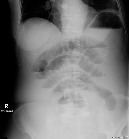
Case courtesy of Dr Craig Hacking, Small bowel obstruction on AXR. Radiopaedia.org. Link
Abdominal X-Ray Interpretation Using PPPE + ABCDO
Step 1: PPPE – Technical Adequacy
-
Projection/Position: Supine AP abdominal radiograph (with an additional erect view). The images are centered on the abdomen, covering from the diaphragm to pelvis.
-
Patient Details: The radiographs are labeled with the correct patient identifiers and date (confirming they belong to our 75-year-old female patient).
-
Positioning: No significant rotation (the spine is aligned midline). The entire abdomen is visible.
-
Exposure: The gas patterns, bony structures, and soft tissue outlines (e.g. psoas margins) can be discerned. The erect film shows lung bases to assess free subdiaphragmatic air.
Step 2: A – Air (Gas Pattern)
-
Normal gas pattern: The stomach bubble is visible. However, instead of normal small amounts of gas in small bowel, there are multiple dilated loops of small bowel in the central abdomen. These loops show stretching of the mucosal folds (valvulae conniventes) all the way across the full width of the bowel, distinguishing them as small intestine. The colon is nearly devoid of gas past the ascending colon, indicating distal obstruction.
-
Abnormal gas: The erect film reveals multiple air–fluid levels in the dilated small bowel loops (visible as air-fluid interfaces at different heights). There is no free air under the diaphragm, so no evidence of perforation.
Step 3: B – Bowel (Location, Size, Wall)
-
The dilated small bowel loops are predominantly central. By the 3-6-9 rule (normal diameter: <3 cm small bowel, <6 cm colon, <9 cm cecum), these loops are clearly abnormal (much >3 cm). They stack in a “step-ladder” appearance on the erect film. The large bowel is collapsed (“empty colon sign”), confirming an obstruction proximal to the colon. There is no obvious visible transition point on the plain film, but the context and collapsed large bowel suggest it is likely at the distal ileum or ileocecal valve (common in adhesional obstruction). The bowel walls are not thickened on X-ray, and there are no pneumatosis (air in the bowel wall) signs.
Step 4: C – Calcifications and Bones
-
Calcifications: No abnormal calcifications are seen. In particular, no gallstones or kidney stones are apparent in the renal or gallbladder areas (most gallstones are radiolucent on AXR, and renal stones if present are not causing these symptoms). The abdominal aorta outline is not notably calcified.
-
Bones: The bony structures (ribs, lumbar spine, pelvis, and hips) show age-appropriate degenerative changes but no acute fractures or lytic lesions. The vertebral alignment is normal. (Always remember to glance at the bones on an abdominal film.)
Step 5: D – Devices and Foreign Bodies
-
Internal devices: No medical devices are present (e.g., no NG tubes, feeding tubes, or surgical clips in view). In this case, an NG tube might be considered for management but none is placed yet.
-
Foreign bodies: No foreign bodies are seen on the X-ray (such as ingested objects or retained surgical instruments). The inguinal regions are clear of any obvious hernia-containing loops on the film.
Step 6: O – Other Findings (Organs & “Oh no!”)
-
Soft tissues / Organs: The liver and spleen outlines are partially visible and not overtly enlarged. Psoas margins are faintly seen, suggesting the exposure is decent. There is no abnormal gas in the biliary tree. No obvious mass is visible.
-
“Oh no!” red flags: The key red flag for bowel perforation which is free intraperitoneal air is absent (no subdiaphragmatic air on the erect film. There is also no evidence of strangulation (no pneumoperitoneum, no gas in the bowel wall, no portal venous gas on this plain film). However, the multiple air-fluid levels and distended loops confirm a mechanical small bowel obstruction.
Summary of Findings and Diagnosis
The abdominal X-rays demonstrate multiple distended loops of small intestine with air-fluid levels, and a collapse of the large bowel, consistent with a Small Bowel Obstruction (SBO). Given the patient’s history of prior abdominal surgery, the most likely cause is adhesions (scar tissue causing obstruction). There are no signs of perforation on the radiograph.
Diagnosis: Adhesional Small Bowel Obstruction.
Key Teaching Points
-
Classic AXR Signs of SBO: Look for centrally located dilated small bowel loops (>3 cm) with valvulae conniventes visible across the entire width of the bowel. On an erect AXR, multiple air-fluid levels stepwise down the abdomen are classic. A “string-of-pearls” sign (small pockets of air between valvulae on an erect film) is another clue to SBO in an obstructed, fluid-filled small intestine. In contrast, the colon will have little to no gas beyond the obstruction.
-
Common Causes: In adults, adhesions from prior surgeries are the #1 cause of SBO. Other causes include hernias, tumors, or strictures.
-
Pitfalls: An ileus (non-mechanical paralysis of bowel) can mimic obstruction on X-ray (multiple dilated loops, but usually both small and large bowel are diffusely distended). Differentiating ileus from true obstruction may require clinical correlation or CT. Also, early high-grade SBO may show little air in loops (if vomited out). A completely gasless abdomen in a symptomatic patient is another “oh no” situation, potentially a closed-loop obstruction.